|
|
|
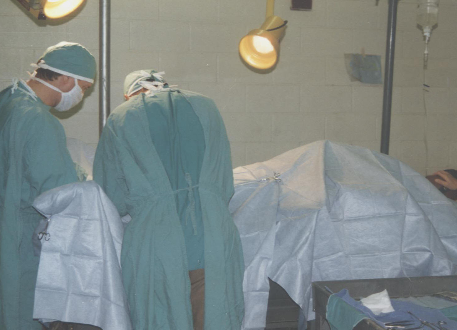
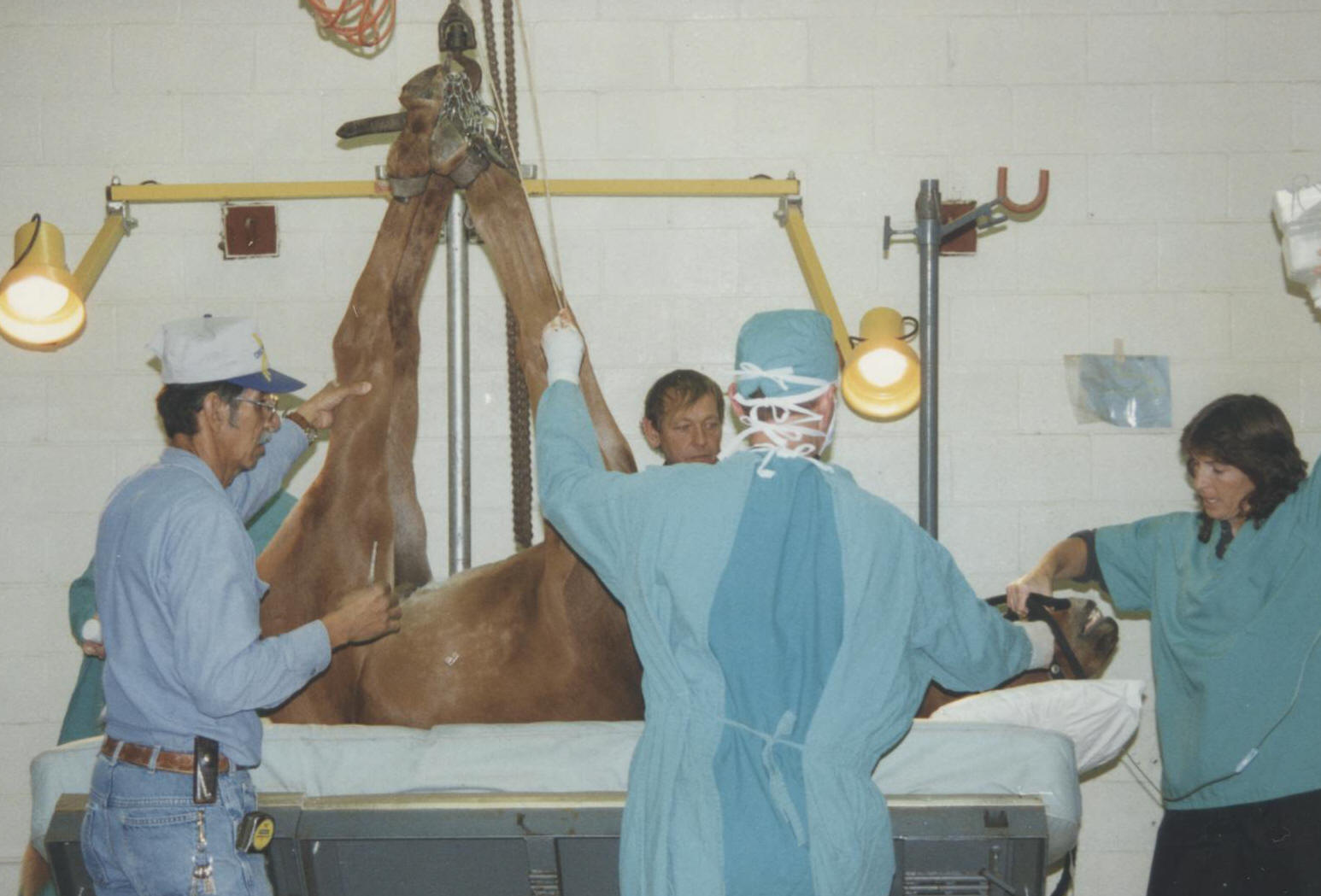
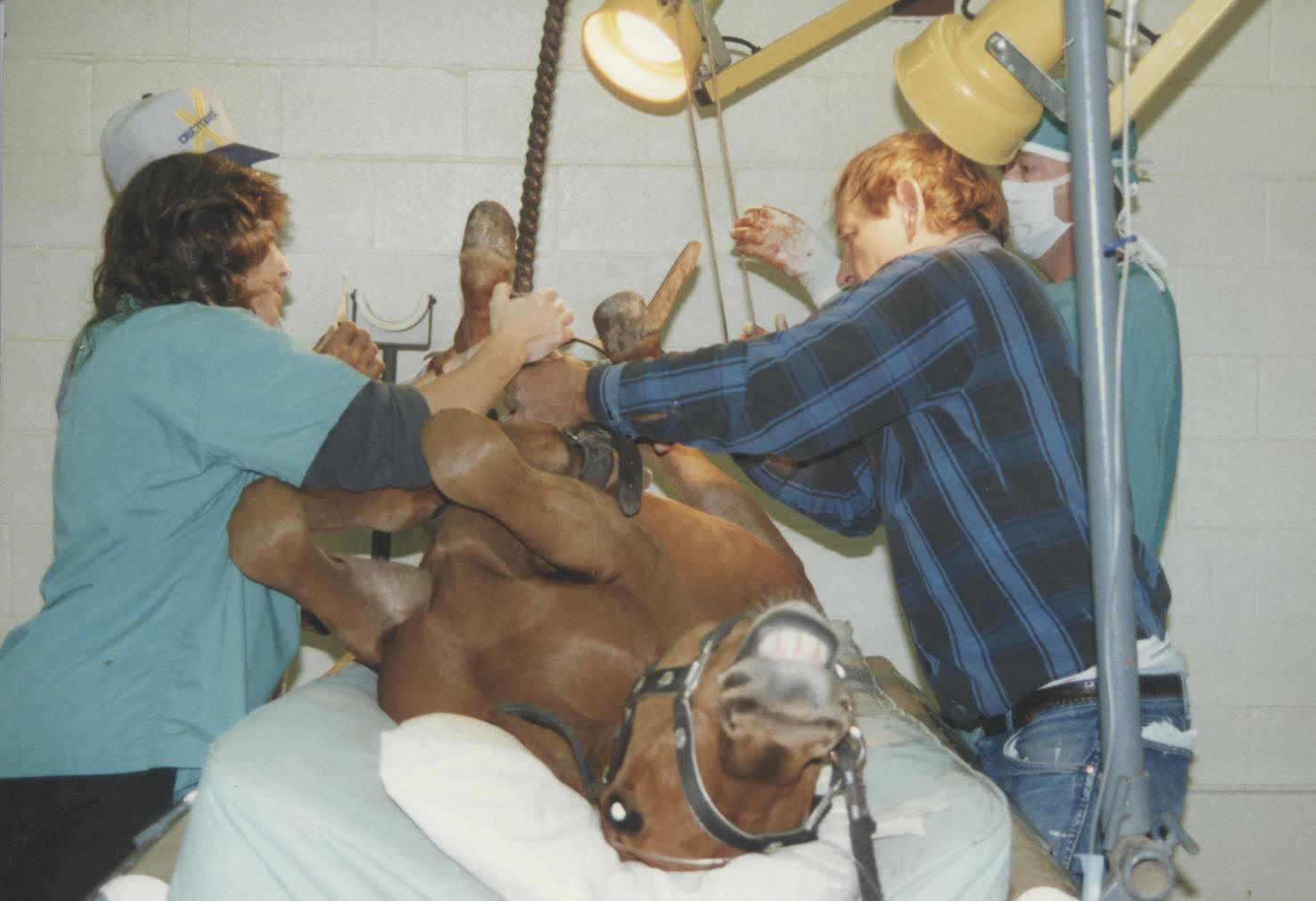
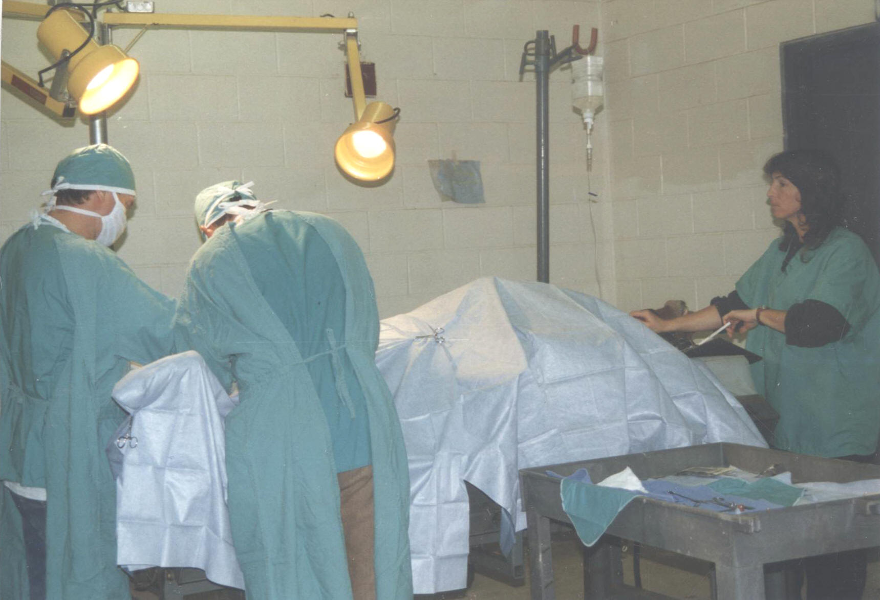
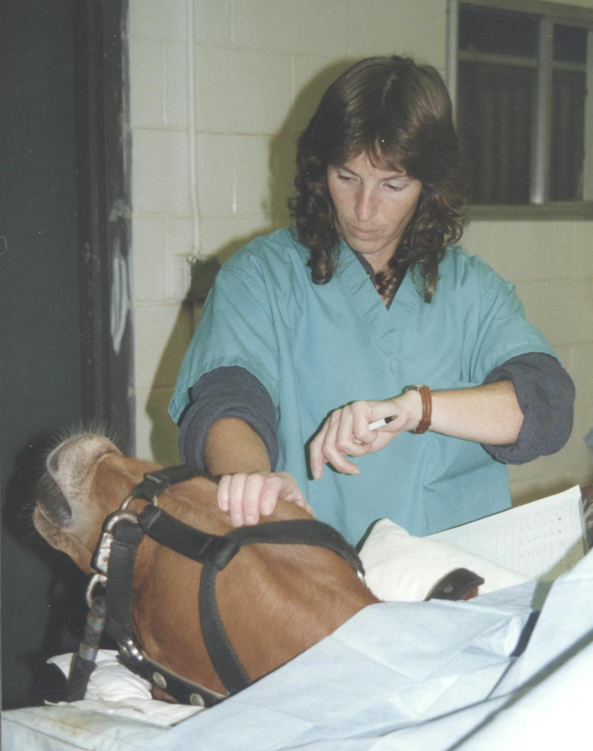
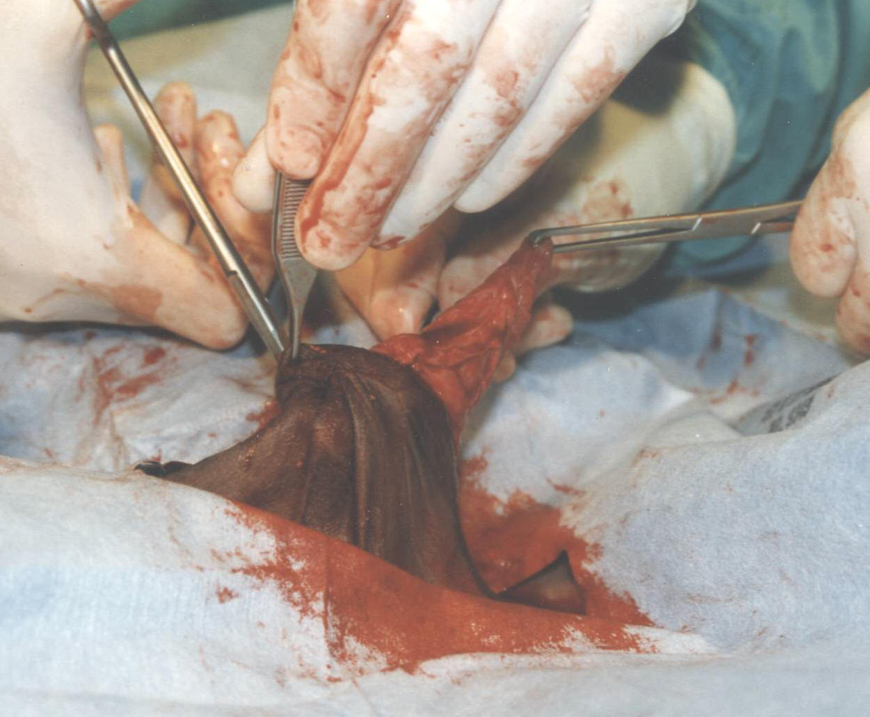
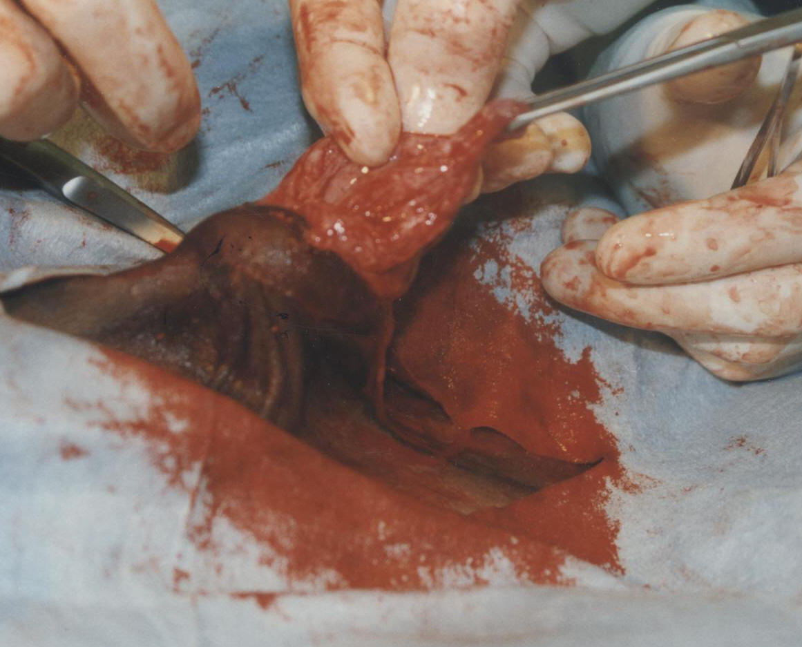
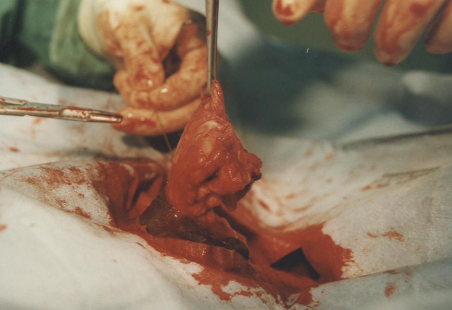
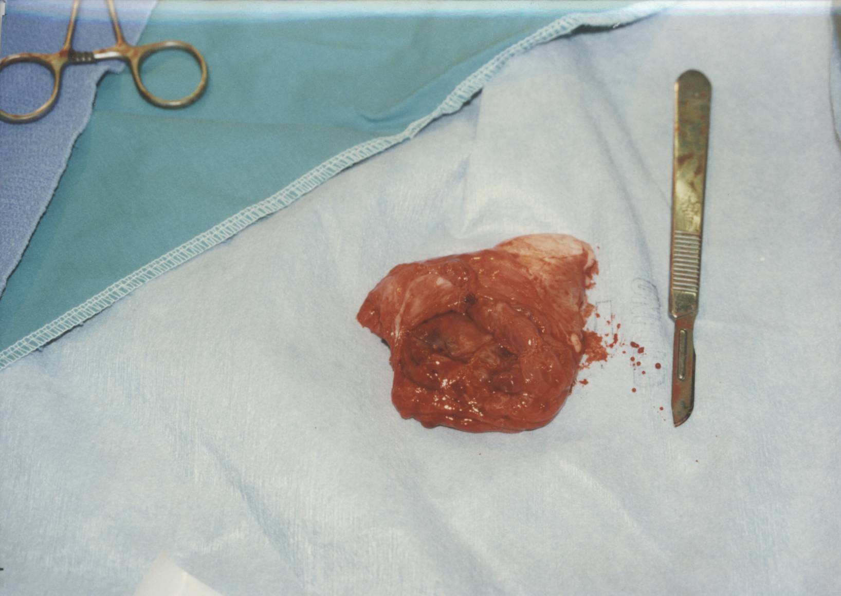
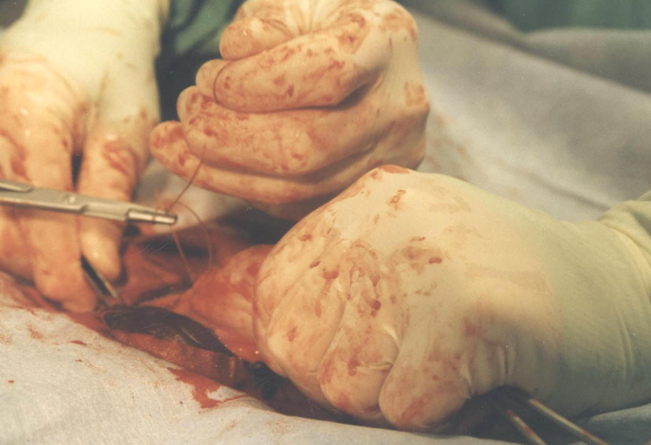
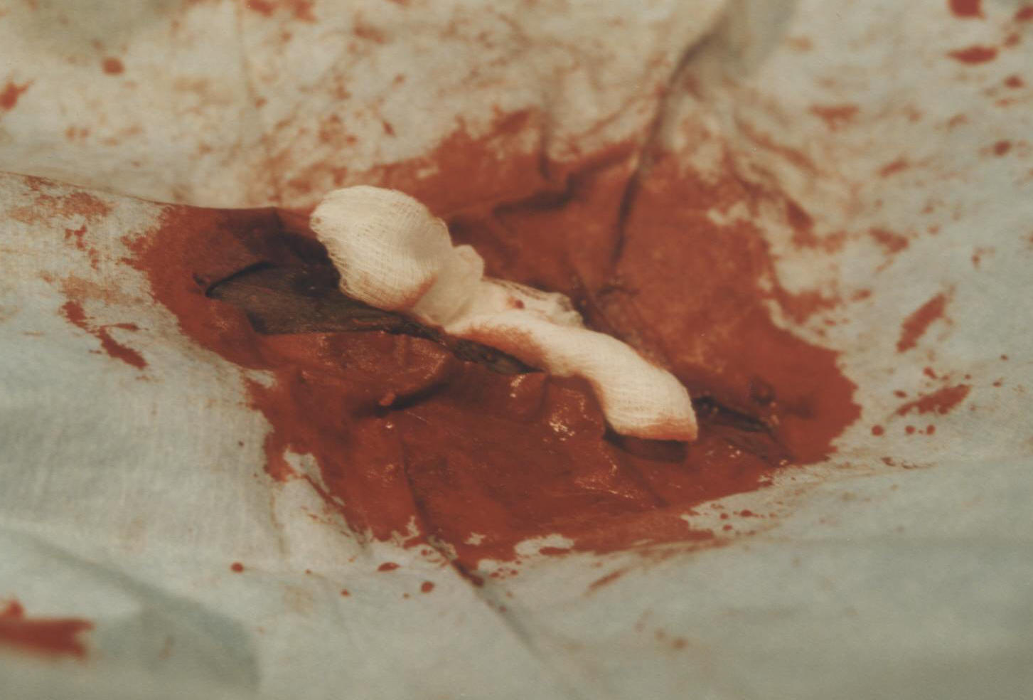
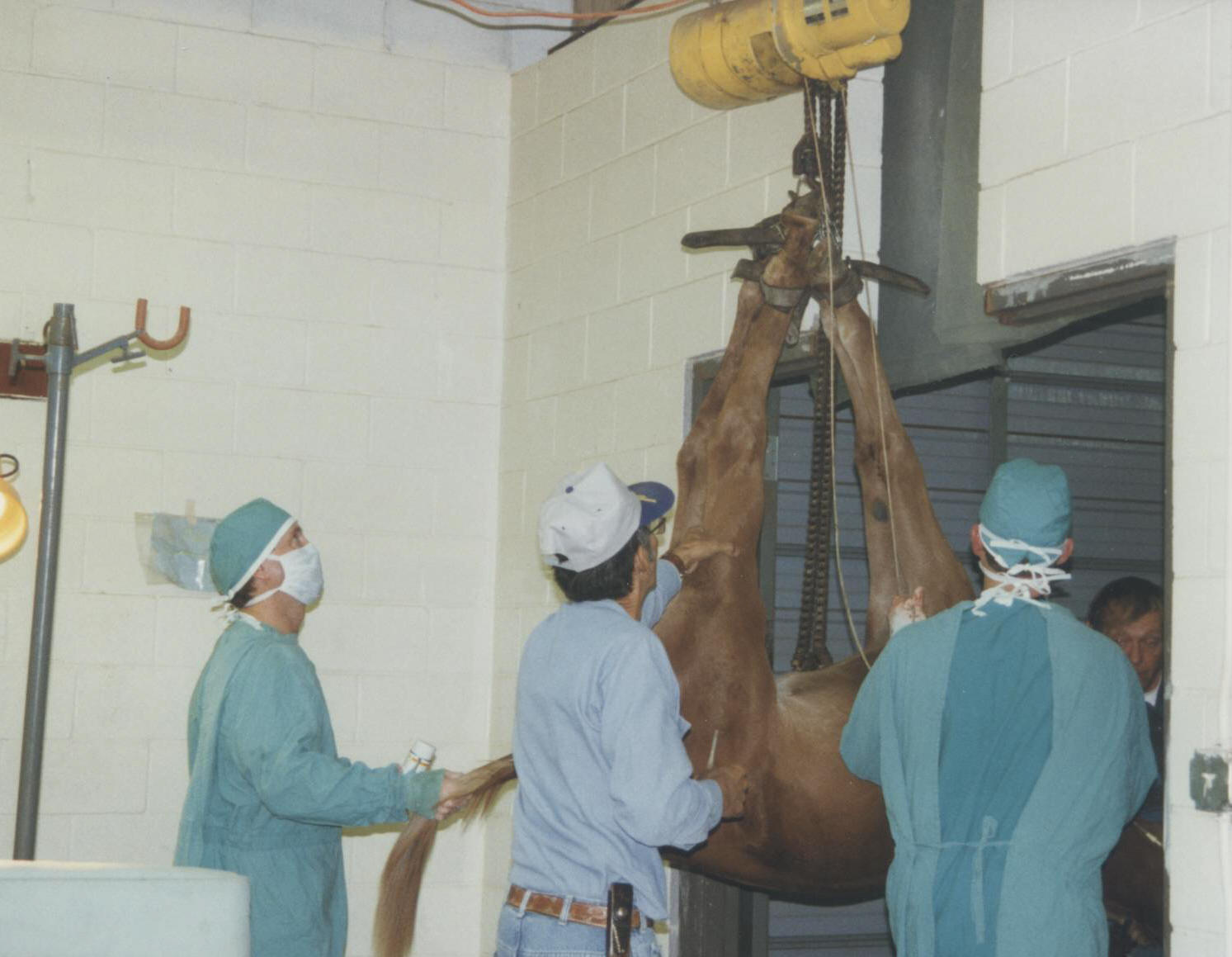
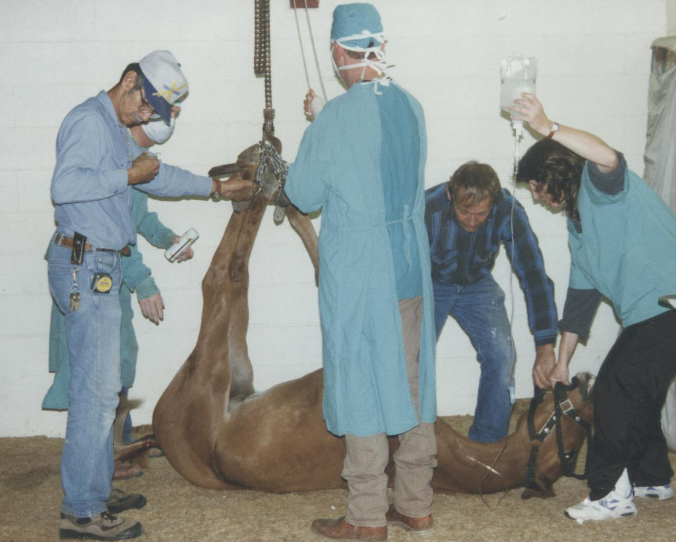
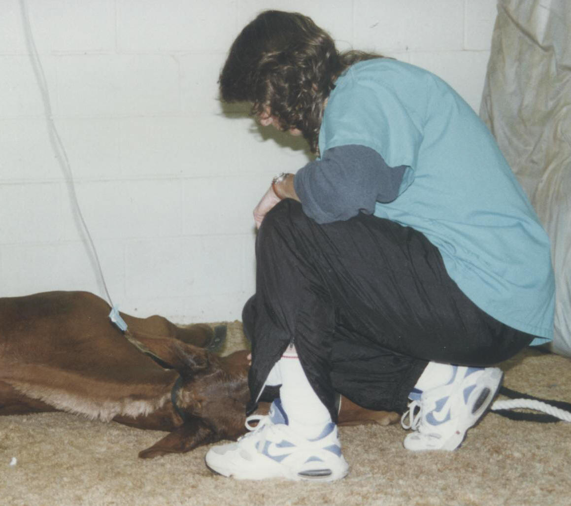
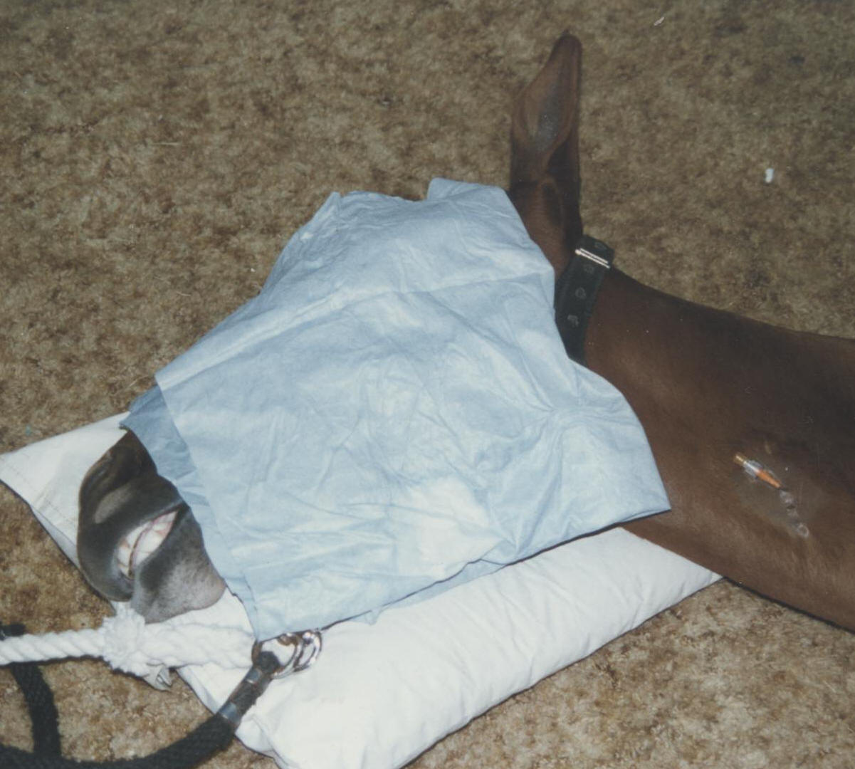
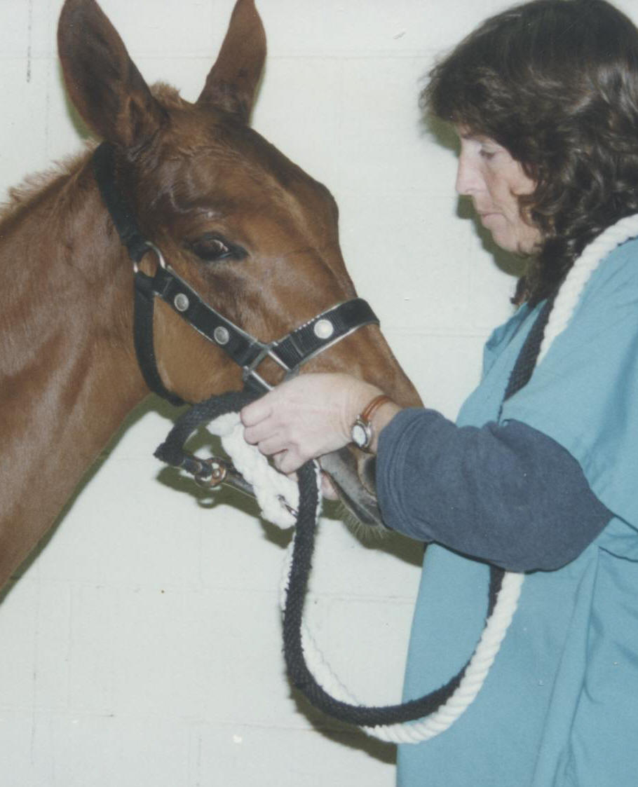
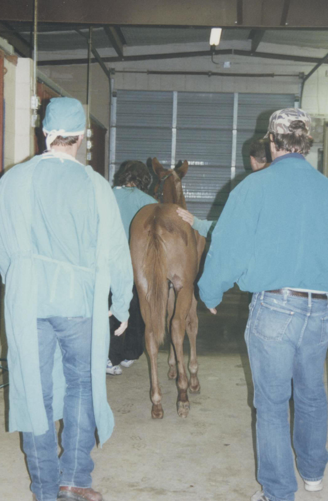
Following a routine castration, a mule colt develops subcutaneous fibrosis/hyperplasia that requires another surgery. Following a routine castration in 1998, one our mule colts, Cowboy Up, developed a growth that gave the appearance that he still had a testicle. The original procedure included the castration of two mule colts. Both mules recovered nicely and showed no signs of problems. Cowboy, however, later developed a pocket that made him look like an unaltered mule. A judge in a weanling gelding class even questioned his being gelded. Palpation revealed a seemingly fibrous mass, the cause of which was unknown. Our only option for Cowboy was exploratory surgery since the nature of the mass was a indeterminate. Of course, for cosmetic reasons we would need to remove the growth, but we had other health concerns for Cowboy. While we were certain that this probably was a simple benign mass, we wanted to rule out a number of other possibilities. The likelihood of the bowel having slipped down the inguinal ring creating a hernia that could later be strangulated was of concern and, of course, we wanted to rule out any infection or abscess. My veterinarian, Dr. Carl Gardner, contacted Tex Taylor to consult on Cowboy=s case. Tex had seen this in more mule colts than horse colts and was familiar with syndrome. Dr. Taylor agreed that it was probably just over reaction of the subcutaneous fat and scar tissue caused by the body=s healing process, but no chances should be taken in case the bowel or infection was involved. Consequently, when Cowboy was prepped for surgery, it was done with full intent to do major surgery if the veterinarians found complications that required more aggressive treatment. What follows is a photo story of Cowboy=s surgery. We were fortunate. The growth was simply fibrous tissue and fat. Cowboy recovered nicely and looks like a gelding again. Thanks again, to Dr. Carl Gardner for his surgical expertise and to Dr. Tex Taylor for his willingness to consult with other veterinarians about the health care of mules and donkeys. |
  |
 |
  |
 |
 |
 |
 |
 |
 |